Calibration method of length of microscope micrometers
Published date and time: 2020-9-17, 15:36
Date and time of modification: 2020-9-17, 15:36
Author: Food-Men
The microscope micrometers are measuring Instruments which are used to measure the length of microorganisms, cells, or other tiny objects. They must be placed in a microscope for measurement. Microscope micrometers are classified into eyepiece micrometers and objective micrometers. The microscope micrometers are commonly used testing equipments and Instruments in the food industry and microbiological research fields. This article introduces the calibration and calculation methods of the length of the microscope micrometers.
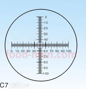
The eyepiece micrometer: is a circular piece of glass which is placed in the eyepiece of the microscope. As shown in the picture of the eyepiece micrometer of microscope, the center is engraved with a precise scale. Usually divide 5mm into 50 grids, and the actual grid is equal to 100μm. The size of the scale changes with the magnification of the eyepiece and objective used. It must be calibrated with the objective micrometer before using.
Picture of the eyepiece micrometer of microscope
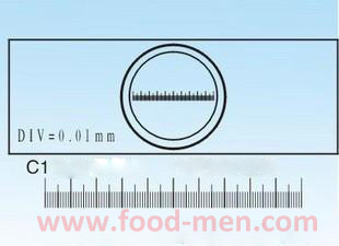
The objective micrometer: is called the stage micrometer. It is a special glass slide that is placed on the stage of microscope. As shown in the microscope objective micrometer picture below, there is a small circle in the center. The circle is engraved with a scale, and the line of 1 mm in length is divided equally into 100 small cells, and each small cell is equal to 10μm. The lower part of the figure, below C1, is an enlarged view of the scale inside the circle. The scale inside the circle is the standard length which is used to calibrate the eyepiece micrometer.
Picture of microscope objective micrometer
Calibration of Microscope Micrometer
(1) Remove the eyepiece tube, unscrew the eye lens on the eyepiece, place the eyepiece micrometer on the middle partition of the eyepiece, make the scale side facing down, then screw the eye lens into the eyepiece tube.
(2) Place the objective micrometer on the stage of the microscope with the scaled side facing up, just like observing the specimen, so that the small circle with the scale is at the center of the field of view.
(3) Firstly, observe with a low magnification eyepiece and focus in. After seeing clearly at the scale of the micrometer of the objective lens, turn the eyepiece so that the scale of the eyepiece micrometer is parallel with the scale of the objective micrometer, and make first line on the left side of these two micrometers coincides. Look to the right for another coincident line.
(4) Record the number of scales of the eyepiece micrometer between the two coincidence lines and the number of scales of the objective micrometer.
Calculation Method of the length of Microscope Micrometer
(1) Eyepiece micrometer length per scale (μm) = two overlapping scales between the objective lens micrometer scale number × 10 / two overlapping scales between the eyepiece micrometer grid number.
(2) In the same way, measure the actual length of each scale on the micrometer is measured under different objective magnifications.
(3) The dimensions of the eyepiece micrometer after the measurement are only applicable to the magnification times of the eyepiece and objective lens of the microscope which are used for the measurement. If their magnification times change when replace the objective or eyepiece, recalibration and calculations must be performed.
——————————————————————
Introduction of related equipment
1. LP-5 Biological Microscope: Uses monocular observation to observe extremely fine microorganisms and has a simple structure.
2. LP-135 General Biological Microscope: Uses binocular observation to observe extremely fine microorganisms.
3. LP-32 Biological Microscope: Has binoculars and a photo lens, which can observe and take pictures of extremely fine microorganisms.
4. XD-1 Inverted Biological Microscope: Can directly observe and photograph the live microorganisms in the petri dish.
5. XD-3 Inverted Biological Microscope: Can directly observe and photograph the live microorganisms in the petri dish.
6. XD-3PMC Inverted Biological Microscope: Can be configured with a polarized modulation phase contrast viewing system. The XD-3PMC inverted biological microscope can directly observe and photograph the live microorganisms in the petri dish.
7. Microscope Slide and Coverslips: Are placed on the microscope stage to facilitate the observation of microorganisms.
——————————————————————